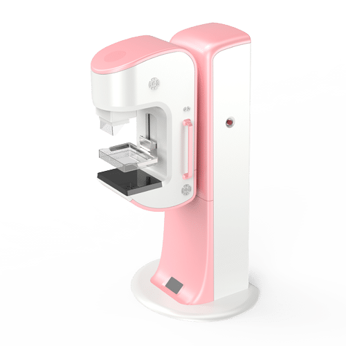
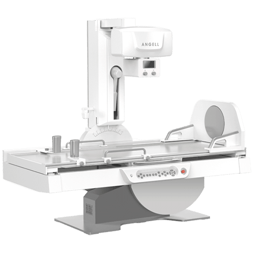
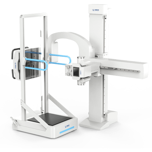
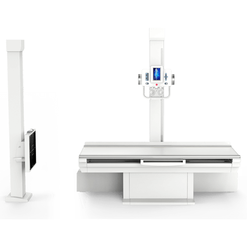
High Frequency Generator
The Floor-mounted DR Series are equipped with a powerful high frequency generator with multiple adjustable settings and reliable performance. Significantly improve the quality of images by adapting the contrast, brightness, density, or sharpness to suit the needs of your patients. In addition to achieving fast interrogation and extinction times typical of high frequency generators, you will also be able to easily control the beam intensityand beam penetration.
A-Si Flat Panel Detector
Our Floor-mounted DR Series fully incorporated the newest a-Si technology in its design, enabling a greater use of computer power with a reduction in hardware cost. Not only is the production of amorphous silicon eco-friendly, but when compared to traditionally used models (like c-Si modules) the a-Si detectors have shown a significant performance improvement. When compared to X-ray film technology, flat panel detectors have manifested greater sensitivity and speed, in addition to maintaining better image quality at a lower dose.
Optional: XG-2 or XG-3
You won’t have to worry about damaging the anodes or tubes in the near future, because both theXG-2 and XG-3 X-ray tubes come with two focal spots – a small one (0.6mm) for procedures that require detailed imaging of smaller regions, and a large one (1.0mm) to conveniently enable a higher heat capacity of the tubes. Both X-ray tubes were specifically designed for a maximally efficient energy conversion for successful low dose examinations whenever possible. For better image quality and faster rotational speed consider our XG-3. With greater heat capacity and more rotations per minute, this DR Series can complete examinations faster while generating images of even better quality.
| Flat Panel Detector |
| Model |
TCQ-III |
| Detector technology |
a-Si Flat Panel Detector |
| Image size |
430mmx430mm |
| Static pixel matrix |
3072×3072 pixels |
| Spatial resolution |
3.7 lp/mm |
| Output grayscale |
16 bit |
| Acquisition time |
≤7 seconds |
| Auto-tracking function |
available |
| Size |
17×17 / 14×17 |
| High Frequency Generator |
| Model |
FSQ-50 |
| Output power |
50kW |
| Frequency |
60kHz |
| kV range |
40 ꟷ 150kV, 1kV step |
| mA range |
100 ꟷ 650mA |
| mAs range |
0.4 ꟷ 650mAs |
| Exposure time |
0.004 ~ 3.2 s |
| Input power |
380V, three phases |
| X-ray Tube |
| Model |
XG-2 |
XG-3 |
| Focal spots |
0.6mm/1.2mm |
0.6mm/1.2mm |
| Voltage |
150kV |
150kV |
| Anode type |
rotating |
rotating |
| Anode heat capacity |
300kHU |
350kHU |
| Anode rotating speed |
2800r/min (50Hz) |
9500r/min (50Hz) |
| Output power |
20kW/50kW |
33kW/78kW |
| Floor-mounted System |
| Model |
PC-3 |
| Bed size |
2100x750x640mm |
| Longitudinal movement range of the bed surface |
±500mm |
| Lateral movement range of the bed surface |
±125 |
| Rotation range of the column |
±180º (lockable at 0º~90º) |
| Vertical movement range of the column along the bed |
±900mm |
| Vertical movement range of the x-ray tube along the column |
450-1850mm |
| Vertical movement range of the bucky stand cassette |
450-1850mm |
| Manual control of bed movement |
available |
| Electrical control of other model systems |
available |
| Collimator |
| Model |
XS-2 |
| Peak tube voltage |
150kVp |
| Power |
150w (24V AC) |
| Auto-close time |
30 seconds |
| Brightness |
≥160lux (100cm) |
| DX Ray Diagnostic Workstation |
| Model |
TX-2 |
| CPU |
Dual-core ≥3.0 |
| RAM |
4GB |
| Hard disk |
500GB |
| Display |
1920×1200 |
| Imaging Part Indicator, Tissue Equalisation, Filter Correction, Grayscale |
available |
| Transform, Window/Level Adjustment, Gamma Correction, ROI Equalisation |
available |
| Black/White Reversion, Image Segmentation, Mark, Enhancement, Smoothing |
available |
| Sharpening, Compression, Magnification Graphic Text Report, Printing |
available |
| Film Printing, Supports standard DICOM 3.0 Laser Film Printer |
available |
| DICOM 3.0 Format, Compatible to transfer to PACS |
available |
